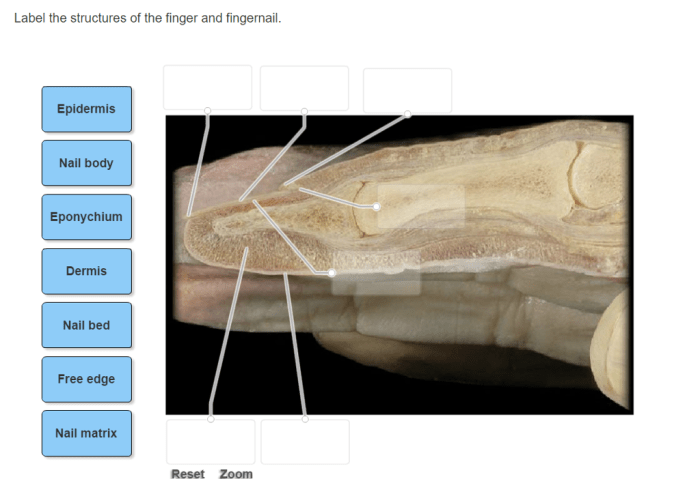
Label the structures of the finger and fingernail. – Introducing the intricate anatomy of the finger and fingernail, this comprehensive guide delves into the structures that orchestrate their remarkable functions. From the finger’s skeletal framework to the fingernail’s protective layers, we unravel the complexities that empower these essential body parts.
Unveiling the intricate arrangement of bones, joints, muscles, and tendons within the finger, we explore the mechanisms that govern its dexterity and range of motion. Simultaneously, we dissect the fingernail’s anatomy, encompassing the nail plate, nail bed, and cuticle, elucidating the processes that contribute to its growth and resilience.
Structures of the Finger: Label The Structures Of The Finger And Fingernail.

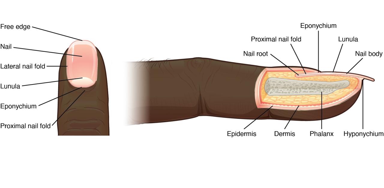
The finger is a complex structure consisting of bones, joints, muscles, and tendons. The bones of the finger are called phalanges and are arranged in three rows: proximal, middle, and distal.
Phalanges
The proximal phalanx is the longest and thickest bone of the finger and connects to the metacarpal bone at the base of the finger. The middle phalanx is shorter and thinner than the proximal phalanx and connects to the proximal phalanx.
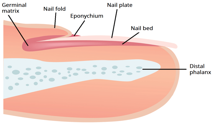
The distal phalanx is the smallest and thinnest bone of the finger and supports the fingernail.
Joints
The metacarpophalangeal joint (MCP joint) is the joint between the metacarpal bone and the proximal phalanx. The interphalangeal joints (IP joints) are the joints between the proximal and middle phalanges, and between the middle and distal phalanges.
Muscles and Tendons, Label the structures of the finger and fingernail.
The muscles of the finger are responsible for flexing and extending the fingers. The tendons are tough bands of tissue that connect the muscles to the bones.
Structure of the Fingernail

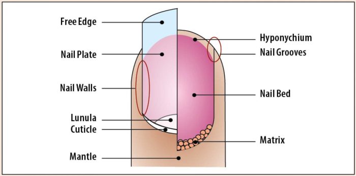
The fingernail is a hard, protective covering that grows from the matrix, which is located at the base of the nail. The nail plate is the visible part of the fingernail and is composed of keratin, a protein that is also found in hair and skin.
Nail Bed
The nail bed is the skin that lies beneath the nail plate. It provides nourishment to the nail and helps to keep it attached to the finger.
Cuticle
The cuticle is a thin layer of skin that surrounds the base of the nail plate. It helps to protect the nail from infection.
Nail Growth
The fingernail grows from the matrix at a rate of about 3 millimeters per month. As the nail grows, it pushes the older nail plate forward. The oldest part of the nail plate is at the free edge of the nail.
Clinical Significance

Nail Disorders
There are a number of common nail disorders, including:
- Onychomycosis: A fungal infection of the nail
- Psoriasis: A skin condition that can affect the nails
- Eczema: A skin condition that can cause the nails to become dry and brittle
- Nail trauma: Damage to the nail caused by an injury
Underlying Health Issues
Some nail conditions can be a sign of an underlying health issue, such as:
- Liver disease
- Kidney disease
- Heart disease
- Thyroid disease
Nail Care and Hygiene
Proper nail care and hygiene is important for maintaining healthy nails. This includes:
- Keeping nails clean and dry
- Trimming nails regularly
- Avoiding biting or picking nails
- Wearing gloves when working with harsh chemicals
FAQ Compilation
What is the function of the metacarpophalangeal joint?
The metacarpophalangeal joint facilitates the bending and straightening motions of the fingers.
What causes nail discoloration?
Nail discoloration can result from various factors, including fungal infections, trauma, or certain medications.
How can I maintain healthy nails?
Regular nail trimming, proper hygiene, and avoiding harsh chemicals can contribute to maintaining healthy nails.
